

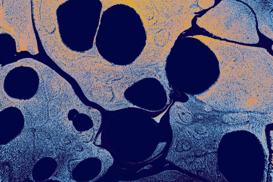

This hidden world within our joints often slips under the radar until pain reminds us it’s there. Cartilage, the silent architect of seamless movement, deteriorates steadily without a single complaint. Yet the secret behind its fading regenerative spark remains a puzzle we’re only now beginning to solve.
As each year passes, the microarchitecture of our joints weathers subtle but cumulative damage, eroding resilience and flexibility. The challenge of restoring lost cartilage extends far beyond simple repair—it demands a profound recreation of its intricate extracellular matrix and the cellular choreography that sustains it. Natural healing paths, robust in youth, wane over time, leaving older individuals vulnerable to stiffness and debilitating pain. Exploring the reasons for this decline could unlock transformative approaches to joint health.
Despite decades of research, engineering a fully functional cartilage substitute remains a formidable task. The load-bearing demands and shock-absorbing qualities of healthy cartilage hinge on a precisely organized matrix of proteins, proteoglycans, and living cells—a blueprint that resists easy imitation. Traditional tissue-engineering prototypes often fall short in replicating the depth of mechanical strength and biological signaling inherent to native cartilage. As a result, true regeneration in aging joints will likely rely on next-generation regenerative medicine strategies that amplify intrinsic healing beyond its natural limits.
Recent open-access work shines a spotlight on a surprising ally in the quest for rejuvenation: macrophages. These versatile cells from the innate immune system orchestrate an elaborate dance between inflammation and repair. Scientists have pinpointed a regulatory gene that steers macrophages toward a pro-regenerative, anti-inflammatory state within cartilage tissue. Unfortunately, expression of this crucial gene declines with age, tipping the balance toward chronic inflammation and compromised maintenance of the cartilage matrix.
Employing cutting-edge single-cell RNA sequencing, researchers compared young and aged animal models following cartilage injury. Their analysis mapped diverse macrophage subpopulations in the joint, revealing a higher abundance of repair-oriented cells in younger specimens. Network analyses then identified Arg-1 (Arginase-1) as a central driver of the beneficial macrophage phenotype. Functional studies confirmed that boosting Arg-1 levels in older animals subdued inflammatory markers and oxidative stress, partially restoring their cartilage repair capacity.
These findings highlight Arg-1 as a promising therapeutic target for enhancing joint resilience in the aging population, setting the stage for innovative treatments that could redefine recovery after cartilage injury. By merging single-cell insights with molecular validation, this research opens a path to counteract the age-related decline in tissue regeneration.
How does aging affect cartilage regeneration and repair?
Cartilage, a resilient connective tissue vital for joint function, shows a markedly diminished regenerative capacity with advancing years. In youth, chondrocytes and extracellular matrix components orchestrate a delicate ballet of maintenance and repair, swiftly responding to minor injuries. However, as the body ages, this choreography falters. The synthesis of key matrix molecules—collagen II and proteoglycans—slows, while the activity of matrix-degrading enzymes accelerates. Microtrauma, once easily resolved, accumulates, paving the way for progressive tissue deterioration and ultimately osteoarthritis.
Underlying this decline is a complex interplay of cellular senescence, reduced growth factor signaling, and chronic low-grade inflammation. Senescent chondrocytes secrete catabolic cytokines and matrix metalloproteinases, tipping the balance toward degradation. At the same time, diminished responsiveness to anabolic signals such as transforming growth factor-β (TGF-β) undermines the tissue’s ability to rebuild. Researchers exploring novel senotherapeutics have found promise in approaches that clear senescent cells or rejuvenate their function. For instance, Rubedo’s early clinical findings for a groundbreaking senotherapeutic treatment highlight how targeted interventions can restore youthful repair mechanisms, offering hope for aged cartilage.
Another challenge lies in recapitulating the cartilage’s complex extracellular matrix architecture in engineered tissues. Unlike softer organs, cartilage must withstand compressive loads and shear forces. Tissue engineering efforts often produce pseudo-tissues that lack the precise mechanical resilience of native cartilage, leaving a gap that demands advanced biomaterials and regenerative medicine strategies. As our understanding deepens, combining cell-based therapies with immunomodulation holds potential to rejuvenate aged cartilage and restore joint health.
What role do macrophages play in cartilage maintenance?
Within the joint microenvironment, macrophages act as sentinels and repair facilitators, modulating both inflammation and tissue remodeling. These innate immune cells exhibit remarkable plasticity, toggling between pro-inflammatory (M1-like) and anti-inflammatory (M2-like) phenotypes in response to local cues. In healthy cartilage, a balanced macrophage presence supports debris clearance and stimulates chondrocyte activity through growth factor release.
Following injury, M1-like macrophages secrete cytokines such as TNF-α, IL-1β, and IL-6 to contain damage and recruit additional immune cells. This initial inflammatory burst is essential, yet if prolonged, it impairs cartilage repair by promoting matrix degradation and chondrocyte apoptosis. Conversely, M2-like macrophages foster resolution of inflammation, secrete anti-inflammatory mediators, and encourage extracellular matrix synthesis. Single-cell analyses now reveal that macrophage activation states in joints exist along a spectrum, with specialized subsets correlating with healing outcomes.
Moreover, synovial macrophages interact with fibroblasts, T cells, and osteoclasts, forming a cellular network that influences joint homeostasis. Disruption of this network—whether by chronic inflammation or age-related changes—can shift the balance toward tissue destruction. Investigations into epoxy oxylipins have shown that these lipid mediators can reprogram macrophages toward anti-inflammatory modes, offering a promising route to quell chronic joint inflammation (exploring epoxy oxylipins).
How does single-cell omics uncover age-dependent macrophage behavior?
Traditional bulk analyses mask the heterogeneity of joint-resident immune cells, but single-cell omics technologies shine a spotlight on individual macrophage states. Single-cell RNA sequencing (scRNA-seq) of young versus aged cartilage injuries reveals stark differences: younger animals harbor a higher proportion of anti-inflammatory macrophage subsets, while older counterparts show enriched pro-inflammatory signatures. This divergence underpins the age-related plateau in repair capacity.
By profiling gene expression at single-cell resolution, researchers identified a regulatory gene—Arginase-1 (Arg-1)—as central to anti-inflammatory macrophage function. In young joints, robust Arg-1 expression drives the conversion of toxic L-arginine metabolites into urea and ornithine, dampening oxidative stress and promoting tissue repair. With age, Arg-1 expression wanes, tipping macrophages toward an inflammatory phenotype that hampers extracellular matrix synthesis and chondrocyte proliferation.
These insights stem from comprehensive network analysis of scRNA-seq data, linking Arg-1 decline to downstream effects on cytokine production and reactive oxygen species release. The study illustrates how age-dependent molecular mechanisms converge on macrophage behavior, providing a mechanistic explanation for reduced cartilage regeneration in older individuals. Armed with this knowledge, scientists can now design targeted interventions to restore youthful macrophage profiles and reinvigorate joint healing.
Why is Arg-1 crucial in macrophage-driven inflammation?
Arginase-1 (Arg-1) serves as a biochemical pivot in macrophage metabolism, controlling the fate of L-arginine and, by extension, the balance between inflammatory and reparative states. In anti-inflammatory macrophages, Arg-1 competes with inducible nitric oxide synthase (iNOS) for L-arginine, reducing nitric oxide production and curbing oxidative damage. The resulting metabolites fuel polyamine synthesis, enhancing cell proliferation and matrix deposition—key processes for effective cartilage repair.
Functional assays demonstrate that overexpressing Arg-1 in aged joints decreases pro-inflammatory cytokines and reactive oxygen species, partially rescuing the impaired healing phenotype. Conversely, Arg-1 deficiency accelerates cartilage breakdown, underscoring its protective role. These findings position Arg-1 not merely as a marker but as a therapeutic target. Interventions that upregulate Arg-1 expression or mimic its enzymatic activity could shift macrophage populations toward pro-regenerative modes.
Parallel avenues of research underscore the broad power of immunometabolism. For instance, senotherapeutic strategies aim to rejuvenate immune cells by clearing senescent elements or modulating metabolic pathways. Interested readers can explore how breathing techniques like the Wim Hof method influence systemic inflammation and may complement Arg-1–focused treatments, fostering an integrated approach to healthy aging.
How can novel therapies target Arg-1 and macrophages to enhance cartilage repair?
Translating mechanistic insights into clinical solutions requires creative therapeutic design. Gene therapy vectors delivering Arg-1 constructs to synovial macrophages could restore youthful enzyme levels, while small-molecule activators may offer a non-invasive alternative. Nanoparticle-based carriers can ensure targeted delivery, minimizing off-target effects and maximizing joint retention.
Immunomodulatory biologics present another frontier. Antibody fragments or cytokine traps that neutralize pro-inflammatory signals in aged joints set the stage for Arg-1–driven repair. Combining these agents with growth factors like TGF-β or insulin-like growth factor 1 (IGF-1) can synergize, promoting chondrocyte proliferation alongside macrophage reprogramming.
Beyond Arg-1, emerging pipelines illustrate the value of systemic health in joint regeneration. Kailera’s upcoming IPO aims to accelerate development in the obesity-treatment arena (Kailera plans IPO), highlighting how metabolic balance influences inflammatory profiles. Similarly, MindImmune’s work on harnessing the immune system against neurodegeneration (Mindimmune secures grant) underscores the potential of immunotherapies across age-related conditions.
Looking ahead, integrating regenerative medicine with lifestyle interventions—nutrient-dense diets, controlled breathing exercises, and targeted senotherapeutics—could offer a multi-pronged strategy for preserving joint health. As research teams refine Arg-1–based modalities, collaboration across fields will be key to restoring the resilience of cartilage and unlocking new horizons in healthy aging.


